Loculated Pleural Effusion / Cnnzfjdh1mk5bm. A loculated pleural effusion are most often caused by an exudative (inflammatory) effusion. An anechoic effusion can be a transudate or exudate (fig. Most pleural effusions, whether free flowing or loculated, are hypoechoic with a sharp echogenic line that delineates the visceral pleura and lung. Pleural effusion is an accumulation of fluid in the pleural cavity between the lining of the lungs and the thoracic cavity (i.e., the visceral and parietal pleurae). Streptokinase appears to improve the resolution of loculated pleural effusions when chest tube drainage fails to achieve symptomatic relief.
A 2 effusions (those with a poor prognosis) occupy more than 50% of the hemithorax, are loculated, and/or are associated with thickening of the parietal pleural. In left heart failure, which results in elevated pressures in the venous system, there is usually some accumulation of fluid in the pleural space. Many medical conditions can lead to it, so even though your pleural effusion may have to be drained, your doctor likely will target. In chf effusions are bilateral and more on right. Surgical thoracostomy tube placement and radiologically guided catheter drainage are standard therapy for loculated pleural fluid collections.
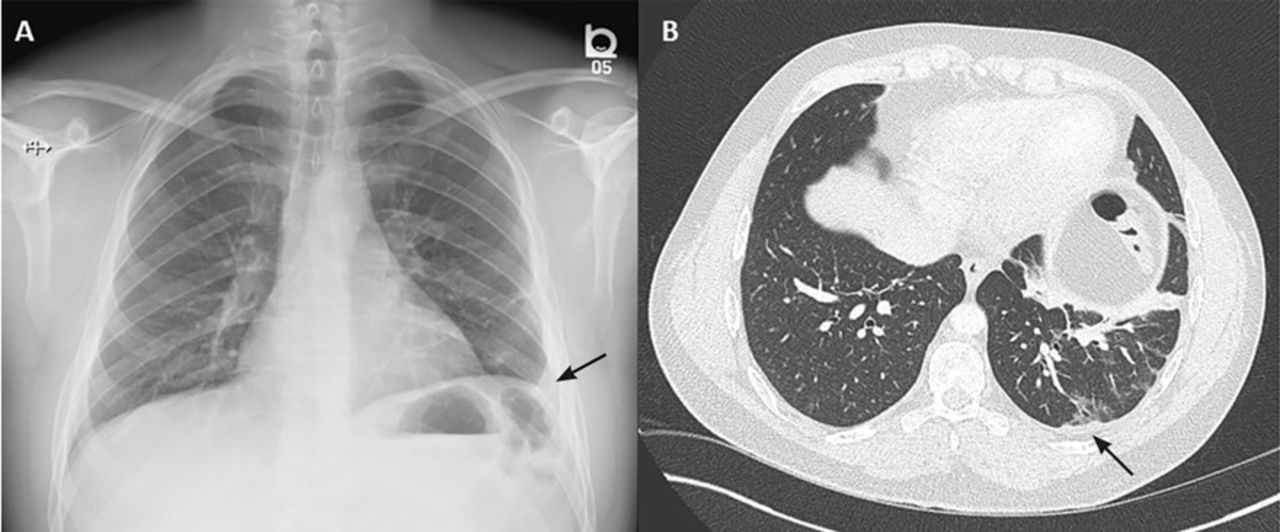
This case highlights the atypical but unique presentation of a transudative pleural effusion and demonstrates the risk of repeated thoracocentesis complicating a simple clinical presentation.
674 views reviewed >2 years ago The fibrinolytic agents, if used early in loculated pleural effusions, break loculations and early pleural peel thereby facilitating pleural space drainage. This scarring, also known as fibrosis, restricts lung function and may cause chest pain and difficulty breathing. Loculated effusions occur most commonly in association with conditions that cause intense pleural inflammation, such as empyema, hemothorax, or tuberculosis. Consider thoracostomy tube drainage if: The largest pocket of fluid is present posteriorly at the right lung base, with associated atelectasis and minor consolidation. An unusual case of vanishing tumor. Normally, a small amount of fluid is present in the pleura. Complex septated, complex nonseptated, or homogeneously echogenic effusions are always exudates (fig. Pleural thickening is a condition triggered by asbestos exposure that causes the pleural lining of the lungs, known as the pleura, to thicken with scar tissue. Causes of an exudative effusion are malignancy, infection, or inflammatory disorders such as rheumatoid arthritis. An anechoic effusion can be a transudate or exudate (fig. Sometimes in the setting of pleuritis, loculation of fluid may occur within the fissures or between the pleural layers (visceral and parietal).
Loculated pleural effusion masquerading as mediastinal tumour had been reported but pleural effusion that conformed to the contour of a lung lobe is rare. Fluid levels in the right and left pleural cavities are often different, known as asymmetrical effusion. 674 views reviewed >2 years ago Pleural effusion is an accumulation of fluid in the pleural cavity between the lining of the lungs and the thoracic cavity (i.e., the visceral and parietal pleurae). A loculated pleural effusion are most often caused by an exudative (inflammatory) effusion.
Tuberculous Pleural Effusion Respiratory Care from rc.rcjournal.com Loculation most commonly occurs with exudative fluid, blood and pus. Treatment may fail if the catheter is not placed optimally within the loculation or if the fluid is hemorrhagic or fibrinous. A pleural effusion is an unusual amount of fluid around the lung. Streptokinase appears to improve the resolution of loculated pleural effusions when chest tube drainage fails to achieve symptomatic relief. Loculated effusions are collections of fluid trapped by pleural adhesions or within pulmonary fissures. Pleural effusions in the intensive care setting. It has been termed vanishing tumor because of its appearance as a mass lesion in the. The pulmonary veins and lymphatics drain the pleural space and return fluid to the heart.
A pleural effusion is an unusual amount of fluid around the lung.
The pleural fluid is called a transudate if it permeates (transudes) into the pleural cavity through the walls of intact pulmonary vessels. In chf effusions are bilateral and more on right. Subcostal loculated pleural effusion in congestive heart failure: In left heart failure, which results in elevated pressures in the venous system, there is usually some accumulation of fluid in the pleural space. Pleural thickening is a condition triggered by asbestos exposure that causes the pleural lining of the lungs, known as the pleura, to thicken with scar tissue. Tell a friend about us, add a link to this page, or visit the webmaster's page for free fun content. Most pleural effusions, whether free flowing or loculated, are hypoechoic with a sharp echogenic line that delineates the visceral pleura and lung. Loculated pleural effusion masquerading as mediastinal tumour had been reported but pleural effusion that conformed to the contour of a lung lobe is rare. Left pleural effusion with high density material at the posterior costophrenic angle. Consider thoracostomy tube drainage if: Loculated malignant effusions however, are inherently resistant to the usual approaches because of nonexpanding underlying lung. Sometimes in the setting of pleuritis, loculation of fluid may occur within the fissures or between the pleural layers (visceral and parietal). The largest pocket of fluid is present posteriorly at the right lung base, with associated atelectasis and minor consolidation.
Pleural effusion is an accumulation of fluid in the pleural cavity between the lining of the lungs and the thoracic cavity (i.e., the visceral and parietal pleurae). The pulmonary veins and lymphatics drain the pleural space and return fluid to the heart. Failure to respond to antibiotic treatment; Pereira w jr, kovnat dm, snider gl. The pleura are thin membranes that line the lungs and the inside of the chest cavity and act to lubricate and facilitate breathing.
Causes of an exudative effusion are malignancy, infection, or inflammatory disorders such as rheumatoid arthritis.
Most effusions start like this and can be easily missed. Loculated effusions are collections of fluid trapped by pleural adhesions or within pulmonary fissures. The pulmonary veins and lymphatics drain the pleural space and return fluid to the heart. Tell a friend about us, add a link to this page, or visit the webmaster's page for free fun content. Complex septated, complex nonseptated, or homogeneously echogenic effusions are always exudates (fig. Pleural effusion that is confined to one or more fixed pockets in the pleural space. Many medical conditions can lead to it, so even though your pleural effusion may have to be drained, your doctor likely will target. Icu patients cannot sit up and the effusion layers posteriorly. Treatment may fail if the catheter is not placed optimally within the loculation or if the fluid is hemorrhagic or fibrinous. An anechoic effusion can be a transudate or exudate (fig. Background tillet and sherry3 were the first ones to use fibrinolytic agents in 1949 in 23 patients who had loculated empyema or hemothorax. The pleural fluid is called a transudate if it permeates (transudes) into the pleural cavity through the walls of intact pulmonary vessels. Subcostal loculated pleural effusion in congestive heart failure:
Share :
Post a Comment
for "Loculated Pleural Effusion / Cnnzfjdh1mk5bm"


Post a Comment for "Loculated Pleural Effusion / Cnnzfjdh1mk5bm"